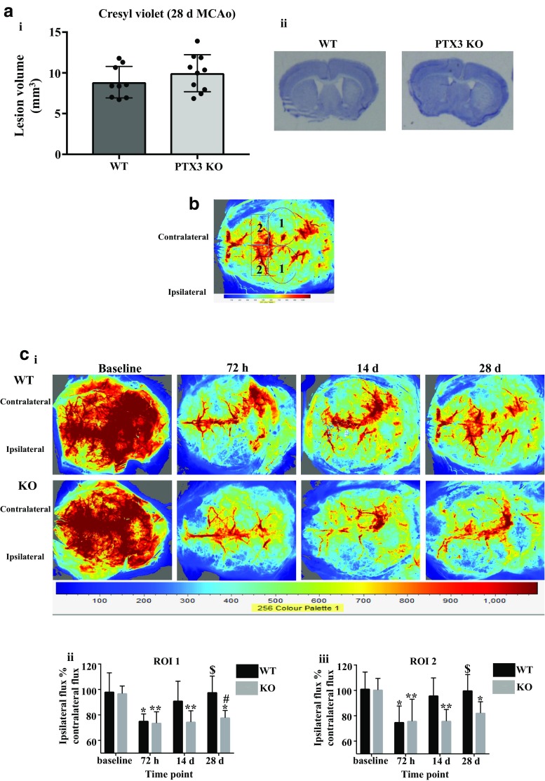
Fig. 1.
PTX3 promotes long-term CBF recovery after cerebral ischaemia. a Infarct volume 28 days after cerebral ischaemia of WT and PTX3 KO mice was assessed on cresyl violet stained brain sections. b Two regions of interest (ROIs) were used to quantify cerebral blood flow (CBF) in WT and PTX3 KO mice as follows; ROI 1 was primary middle cerebral artery (MCA) area and ROI 2 was frontal cortical region. Labels 1 correspond to ROI 1, labels 2 correspond to ROI 2. c i LSCI images at baseline, and 72 h, 14 days, and 28 days post-stroke in WT and PTX3 KO mice. c ii–iii CBF quantified with moorFLPI2 Full-Field Laser Perfusion Imager Review V5.0 software and expressed as ipsilateral flux as % of contralateral flux in two ROIs. Statistical analyses were performed using a unpaired Student’s t test (ns P > 0.05) and c linear mixed modelling followed by Holm-Sidak post hoc (ns P > 0.05, * P ≤ 0.05, ** P ≤ 0.01, *** P ≤ 0.001). * represents significance within genotype from baseline; $ represents significance within genotype from 72 h; # represents significance between genotype. All data are presented as mean ± SD (n = 6–7 per group)

