Fig. 6.

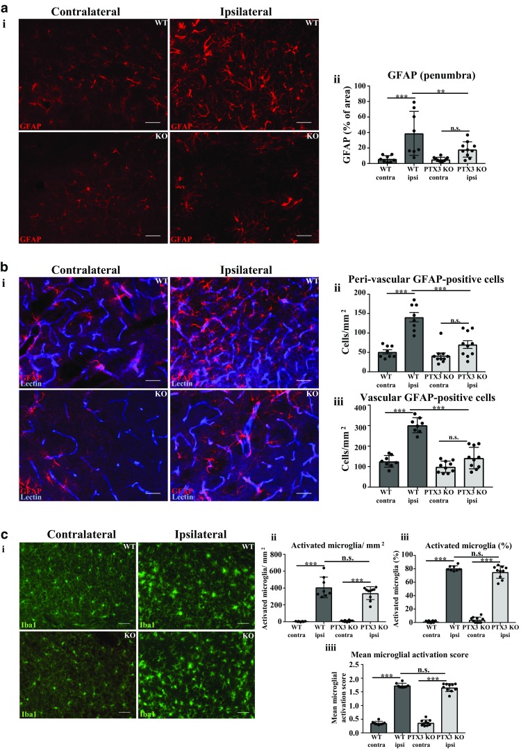
Pentraxin 3 knockout (PTX3 KO) mice express reduced number of vascular and peri-vascular GFAP-positive astrocytes. a i Glial fibrillary acidic protein (GFAP)-positive astrocytes (red) in ipsilateral or contralateral hemispheres of penumbra region in WT or PTX3 KO mice as labelled. Scale bar 50 μM. b i GFAP (red) and lectin (blue) co-immunohistochemistry of ipsilateral or contralateral hemispheres of penumbra region in WT or PTX3 KO mice as labelled. Scale bar 50 μM. c i Iba1 (green) expressing microglia in ipsilateral or contralateral hemispheres of penumbra region in WT or PTX3 KO mice as labelled. Scale bar 50 μM. a ii Quantification of GFAP percentage (%) area staining in ipsilateral or contralateral hemispheres of penumbra region in WT or PTX3 KO mice. b ii Peri-vascular or b iii vascular GFAP+ astrocytes/mm2 in ipsilateral or contralateral hemispheres of penumbra region in WT or PTX3 KO mice were counted with ImageJ software. The c ii number/mm2 or c iii % of Iba1 expressing microglia was evaluated with ImageJ software. c iiii Mean microglial activation score of Iba1 stained microglia was also calculated. Statistical analyses performed using repeated measures two-way ANOVA followed by Sidak corrected post hoc analysis (ns P > 0.05, ** P ≤ 0.01, *** P ≤ 0.001). All data expressed as mean ± SD (n = 8–10)
