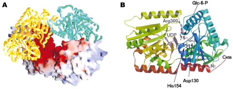
Figure 5.
Crystal structure of trehalose-6-phosphate synthase (OtsA) provided by Gibson et al. [185]. (A) Tetramer of the enzyme. The electrostatic surface representation (positive, blue; negative, red) and the protein cartoon figure are mixed. (B) Ribbon representation of the structure of OtsA. The two ligands, glucose-6-phosphate and UDP, are shown in ball-and-stick model. Figure courtesy of Professor G. J. Davies and Cell Press.

