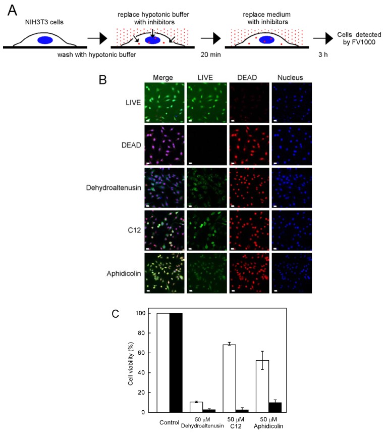
Figure 4.
Inhibitors were incorporated into cells using a hypotonic shift. (A) Scheme of the procedure for a hypotonic shift. (B) NIH3T3 cells incubated for 20 min in hypotonic buffer without or with 50 μM dehydroaltenusin, C12 or aphidicolin. The culture medium was then replaced with medium including 50 μM dehydroaltenusin, C12 or aphidicolin for 3 h. Cells were detected using a LIVE/DEAD® Viability/Cytotoxicity Kit. Red and green indicate dead and live cells, respectively. Dead cells were treated with 70% methanol for 30 min. (C) Cell viability using a hypotonic shift (closed bar) or not (open bar) is shown. Live and dead cells were individually counted from at least 200 cells (from each condition). Control was taken as 100%. Values are shown as the means ± S.E. for four independent experiments. All bars indicate 20 μm.

