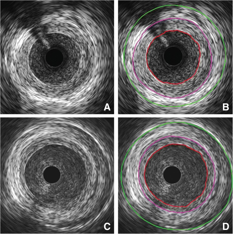
Fig. 3.

Within-patient comparison of intimal hyperplasia using IVUS. Segment of a nonstented SVG to the first obtuse marginal 4.5 years after implantation without (a) and with (b) marking of the lumen (red), EEM (purple) and outer vessel (green). Segment of externally stented SVG to the second obtuse marginal 4.5 years after implantation without (c) and with (d) marking of the lumen (red), EEM (purple) and stent (green)
