Fig. 2.
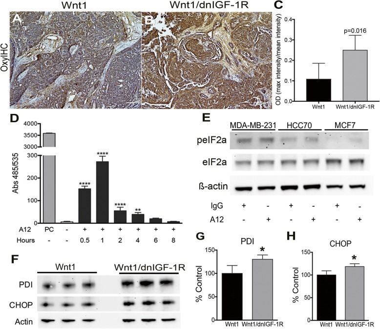
Cellular stress is increased in response to reduced insulin-like growth factor type1 receptor (IGF-1R) function. a, b Representative images showing OxyIHC-stained sections from Wnt1 (a) or Wnt1/dnIGF-1R (b) tumors. c Quantification of OxyIHC-stained sections by 3,3′-diaminobenzidine intensity measurement (n = 5 tumor sections per genotype) (Student’s t test, P = 0.016). d The 2′,7′-dichlorofluorescin diacetate assay analysis of untreated or A12 treated MCF7 cells at subsequent time points: 0.5, 1, 2, 4, 6, and 8 h. PC (tert-butyl hydrogen peroxide positive control; 100 μM) (one-way analysis of variance, untreated versus A12: **P < 0.01, **** P < 0.0001; n = 3, with three technical replicates per experiment). e Representative western immunoblot showing levels of phospho-eukaryotic initiation factor 2-alpha (eIF2α) and total eIF2α protein compared to loading control (ß-actin) in untreated (IgG) and human breast cancer cell lines (MDA-MB-231, HCC70, MCF7) treated with A12 for 24 h. f, h Western immunoblotting showing levels of protein disulfide isomerase (PDI) and C/EBP homologous protein (CHOP) in Wnt1 and Wnt1/dnIGF-1R tumors (f). Densitometry analysis of PDI (g) and CHOP (h) protein expression normalized to ß-actin in Wnt1 and Wnt1/dnIGF-1R tumors. (Student’s t test, *P < 0.05; n = 3)

