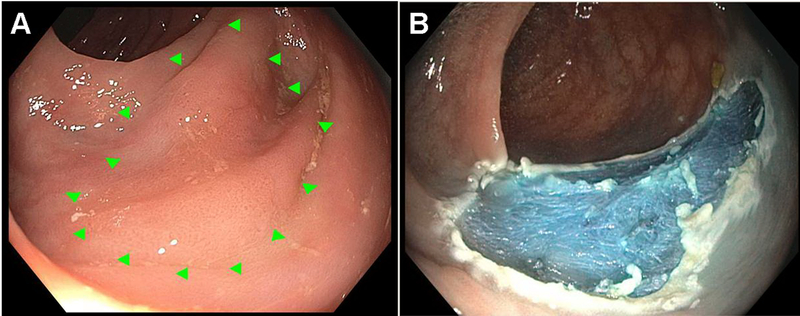
Figure 2:
Example of a 20mm flat (Paris 0-IIb, NICE 2) non-granular laterally spreading lesion referred to a specialized center for EMR. Referring physician described location of polyp in detail, provided high quality endoscopic images (A), avoided biopsy or partial removal during index procedure, and tattooed the opposite wall of the colon to aid the EMR practitioner in locating this subtle polyp. EMR performed 1 month later with excellent results. Histopathology revealed a tubular adenoma without high-grade dysplasia.

