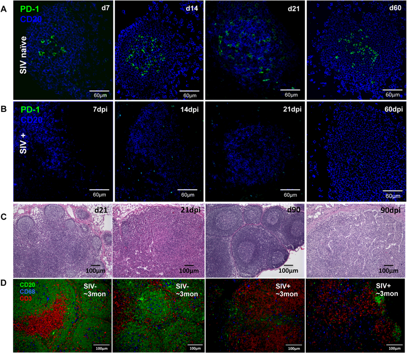
Figure 3. Effects of SIV infection on follicle structure of lymph nodes in neonatal macaques infected with SIVmac251.
Confocal microscopy of PD-1+ cells in follicles of lymph nodes in SIV naïve age-matched (A) and SIV-infected neonates post SIV infection (B). CD20, blue; PD-1, green. (C) Histopathological examination of follicles in the lymph nodes of infants with or without SIV infection at birth. The numbers of follicles were decreased and remaining of follicles were primary (lacked germinal center formation) in SIV-infected infants at 21 and 90 dpi, compared with age-matched controls. Severe lymphoid depletion and impaired follicle formation observed throughout SIV infection in infants. All photomicrographs were taken from H&E stained slides at an original magnification of 100×. (D) Confocal image analysis of B/T-cell and macrophages in lymph nodes in SIV naïve or SIV-infected infants with ~3 months age, as shown CD20 (green), CD3 (red) positive cells and macrophage (blue).

