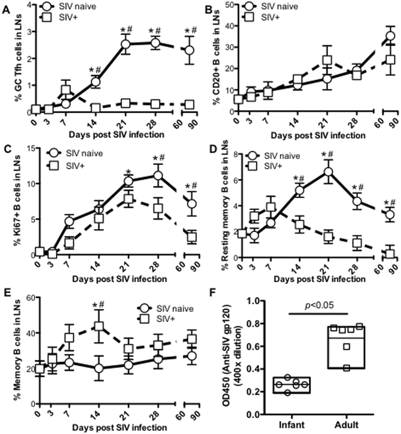
Figure 4. Effects of SIV infection on GC Tfh cell and B-cell differentiation in neonatal macaques infected with SIVmac251.
(A) Changes in GC Tfh cells (CXCR5+PD-1high CD4+ T-cell) in lymph nodes of infants infected with SIV at birth and examined at day 3 (n=3), 7 (n=3), 14 (n=6), 21 (n=5), 28 (n=3) and 2~3 months (n=8) post SIV infection, compared with age-matched uninfected infants (n=35); (B) Percentage of CD20+ B cells in lymph nodes of infants post SIV infection; (C) Proliferation of B cells in lymph nodes of neonates post SIV infection; and (D and E) changes of resting memory B-cell (IgD+CD27+) and memory B-cell in lymph nodes of infants post SIV infection; (F) Levels of plasma anti-SIV gp120 in infants and adults after 3 months SIV infection. *,# p<0.05, compared with newborn (*) or age-matched normal infants (#).

