
Fig. 1.
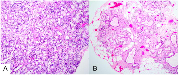
Histopathology of minor labial salivary glands. The sections are from biopsies of a 28-year-old woman (A) and a 65-year-old woman (B), shown at the same magnification. Neither had Sjögren syndrome. (A) This histopathologic section shows normal tissue, with confluent mucous acini and normal-sized intralobular ducts. (B) In contrast, this section shows extensive acinar loss, interstitial fibrosis, ductal dilatation, and fatty replacement. These changes are often seen to varying degrees in older patients (H&E stain, original magnification ×100).