
Fig. 3.
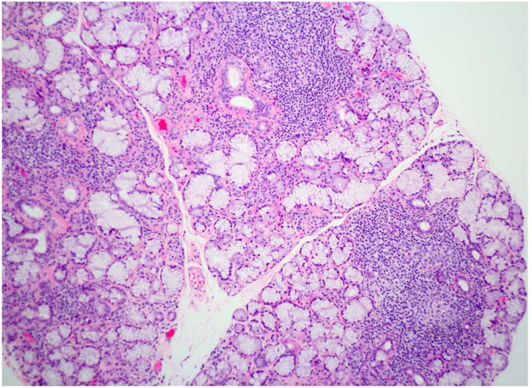
Focal lymphocytic sialadenitis. This section of a labial minor salivary gland biopsy shows the typical features of focal lymphocytic sialadenitis. Note the tightly aggregated lymphocytes surrounding ducts and adjacent to normal-appearing mucous acini. At least 3 foci are evident (H&E stain, original magnification ×100).