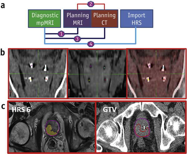
Figure 6: Workflow for incorporation of HRS in BLaStM patient RT planning.
(a) Schematic of the registrations utilized and the migration of the HRS structures: (1) Using prostate anatomical matching, the diagnostic mpMRI is registered with the planning MRI; (2) The planning MRI is fused to the planning CT, using fiducial matching; (3) Using (1) and (2), the diagnostic mpMRI generated HRS contours are migrated to the planning CT; (b) Registration of the planning CT (left) and planning MRI (center) using fiducial matching and the alignment (right); (c) The final result for one patient is displayed where the HRS 6 contour has been migrated to the planning CT using the methods described.

