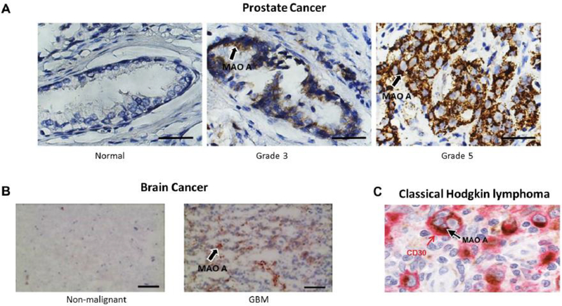
Fig. 5. Elevated expression of MAO A in various human cancers.
(A) High Gleason grade prostate cancers associated with increased MAO A expression. Normal prostatic epithelium and Gleason grade 3 and 5 prostate cancer specimens from a tissue microarray were stained for MAO A. Magnification, 400x; bars, 20 μm. (Adapted from Wu et al., 2014) (B) MAO A expression is increased in human glioma. Non-malignant brain and glioma tissue specimens. Scale bars represent 100 μm (Adapted from Kushal et al., 2016) (C) MAO A is highly expressed by Hodgkin/Reed–Sternberg (HRS) cells of classical Hodgkin lymphoma (cHL). MAO A was detected by immunohistochemistry on formalin-fixed, paraffin-embedded (FFPE) patient material. Double staining for CD30 (red) and MAO A (brown) confirms the localization of MAO A in HRS cells (original magnification 1000×) (Adapted from Li et al., 2017)

