Fig. 3.
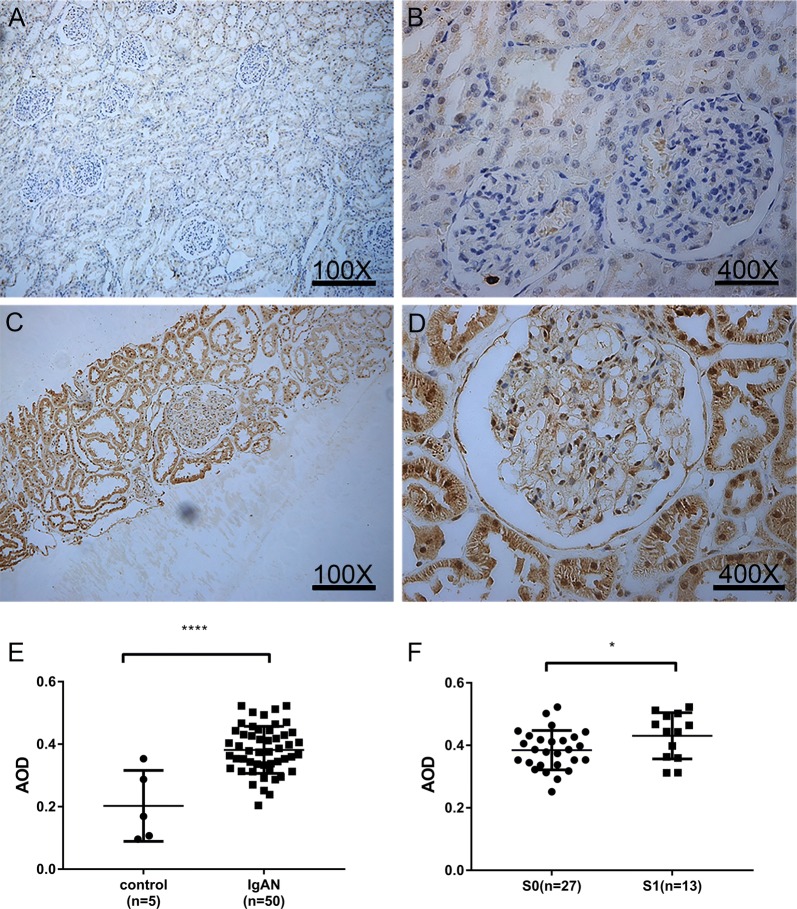
Tissue NLRC5 expression was different between IgAN patients and controls. Expression of NLRC5 in renal tissue of controls (A, B) and IgAN patients (C, D) by immunohistochemistry (A, C 100×), (B, D, 400×). E Relative quantitative analysis of renal tissue NLRC5 expression in controls and IgAN patients, the average optical density (AOD) was calculated by Image-pro Plus 6.0 immunohistochemical analysis software. F Quantitative analysis of renal tissue NLRC5 expression in S0 and S1 groups of IgAN patients according to Oxford classification. E, F by Unpaired t test. *P < 0.05, ****P < 0.0001

