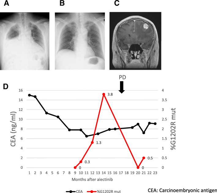
Fig. 3.
Case #1. (a) Chest radiography showing PD 23 months after treatment with crizotinib, with pericardial effusion and right pleural effusion. (b) Chest radiography 13 months after treatment with alectinib. Imaging revealed a moderate response in pericardial effusion and right pleural effusion. (c) MRI imaging showing PD (brain metastasis) 17 months after treatment with alectinib. (d) Graph shows the change of % G1202R mut in cfDNA using ddPCR and CEA levels during the clinical course after treatment with alectinib. % G1202R mut ×100. PD = Progressive disease

