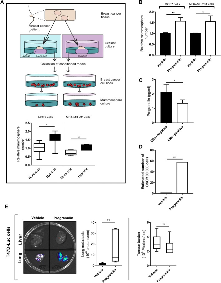
Fig. 1.
Identification of progranulin as a secreted component that influences breast cancer stem cell propagation. a Schematic of the experimental procedure involving ERα-positive primary tumour explants (n = 7) and mammosphere formation of breast cancer cell lines MCF7 and MDA-MB 231 in response to pre-treatment with conditioned media. Results are expressed as relative mammosphere formation (n = 7). *p < 0.05, **p < 0.01 and ***p < 0.001 as calculated by a Student’s t test. b ERα-positive MCF7 and ERα-negative MDA-MB 231 cell lines were treated with 1 μg/ml progranulin for 48 h and then assessed for mammosphere-forming capacity. Results are expressed as relative mammosphere numbers ± SD (n = 3). *p < 0.05, **p < 0.01 and as calculated by a Student’s t test. c Culture media collected from ERα-positive MCF7, T47D and ERα-negative MDA-MB 231 and MDA-MB 468 cultures where analysed for progranulin secretion using human progranulin ELISA (n = 3). *As calculated by a Student’s t test. d ERα-positive MCF7 cells were pre-treated with 1 μg/ml progranulin for 48 h and then injected into NOD SCID gamma mice in serial dilution format. Xenograft results were calculated at day 59 using extreme limiting dilution analysis (ELDA) software to determine the CSC frequency and significance. *p < 0.05, **p < 0.01 and ***p < 0.001. e T47D-luc xenografts where treated with either vehicle (PBS) or 8 μg of progranulin three times per week for 6 weeks by subcutaneous injection. Tumour burden and lung metastases luciferase measurements at the experimental endpoint are expressed as mean photons/second (right, top and bottom respectively) (n = 6). Mann-Whitney U test was used for statistics. **p < 0.01. CSC cancer stem cell, ERα estrogen receptor alpha

