Fig. 2.
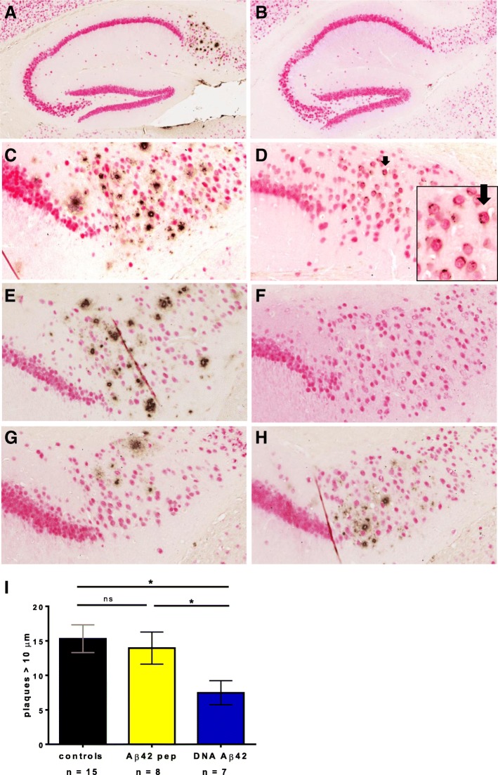
Amyloid-β (Aβ) immunization results in removal of amyloid plaques in brains of triple-transgenic Alzheimer’s disease (3xTg-AD) mice. Brain sections of mice aged 18 months (c and d) and 20 months (a, b, e–h) were stained with a NeuN antibody (red) to detect neurons and an anti-Aβ antibody (McSA1, brown) to detect numerous plaques in the subiculum of the hippocampus in 3xTg-AD mice. a The hippocampus of a 20-month-old female control 3xTg-AD mouse with numerous amyloid plaques is shown (5× magnification). b Hippocampus of a 20-month-old male control 3xTg-AD mouse showing no plaque pathology. c The subiculum of an 18-month-old female control 3xTg-AD mouse is shown at higher magnification (20×). d Aβ staining in the subiculum of an 18-month-old male mouse. Only intraneuronal Aβ can be detected (indicated with arrow and shown at higher magnification in inset). e Numerous plaques in the hippocampus of an untreated 20-month-old 3xTg-AD female mouse. f No plaques were found in 20-month-old wild-type mice. Both immunization regimens, Aβ42 peptide (g) and DNA Aβ42 (h), led to a reduction of plaques in 20-month-old 3xTg-AD mice compared with the control mouse (e). i Images were counted for plaques ≥ 10 μm in a 1-mm2 area of the subiculum/CA1 of the hippocampus by two blinded experimenters. Blue bars show plaque count in DNA Aβ42 trimer-immunized mice (n = 7), and yellow bars show plaque count found in brains of Aβ42 peptide-immunized mice (n = 8). Black bars show the numbers found in age- and gender-matched 3xTg-AD control mice (n = 15). * indicates p value of ≤ 0.05 (unpaired Student's t test)

