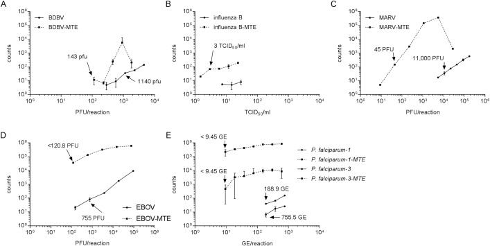
Fig 1. Improved PCR detection following MTE.
BDBV (A), influenza B strain 38 (B), MARV (C), EBOV (D), and P. falciparum (E) nucleic acid were serially diluted. One dilution series was amplified by MTE prior to being run in the SYBR Green assay. The amount of pathogen-specific nucleic acid was determined by real-time PCR using primers internal to the MTE amplification primers. Fig B is shown as a dilution factor because influenza B strain 38 did not titer. Samples were run in triplicate, and the error bars represent the standard deviation of the mean. A two-way ANOVA with Bonferroni determined all dilutions for all viruses tested were significantly different with the exception of the highest influenza B virus concentration (~103 PFU/rxn).

