Fig. 1.
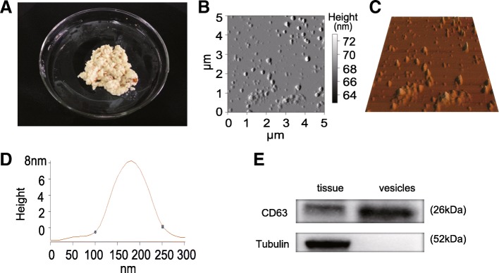
Identification of exosomes in PM. a The morphology of PM. b 2D AFM image of isolated exosome vesicles in PM. c 3D AFM image of isolated exosome vesicles in PM. d The line profile of AFM image for a PM exosome vesicle. X- and Y-axes are the width and height of PM exosome vesicle, respectively. e The expression of CD63 and Tubulin in the crop tissue and exosome vesicles was determined by Western blotting. It can be seen that the isolated exosome vesicles are positive for CD63 but negative for Tubulin

