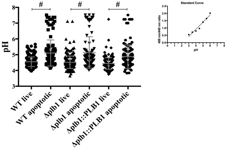
Fig 11. Ph measurment of Cn-containing phagolysosomes.
BMDM infected with WT, Δplb1 and Δplb1::PLB showed no significant difference between the different strains, but there was a significant difference when we compare live versus apoptotic BMDM between the BMDM infected with the different strains. The pH of Cn-containing phagolysosomes was less acidic in BMDM positive for annexin V in comparison to BMDM negative for annexin V. Determination of pH by using a standard curve of buffers with different pH in the presence of nigericin to equilibrate the extracellular and intracellular pH. One-way ANOVA with Dunnett’s multiple comparison test *p > 0.0332, **p < 0.0021, ***p < 0.0002 and #p < 0.0001.

