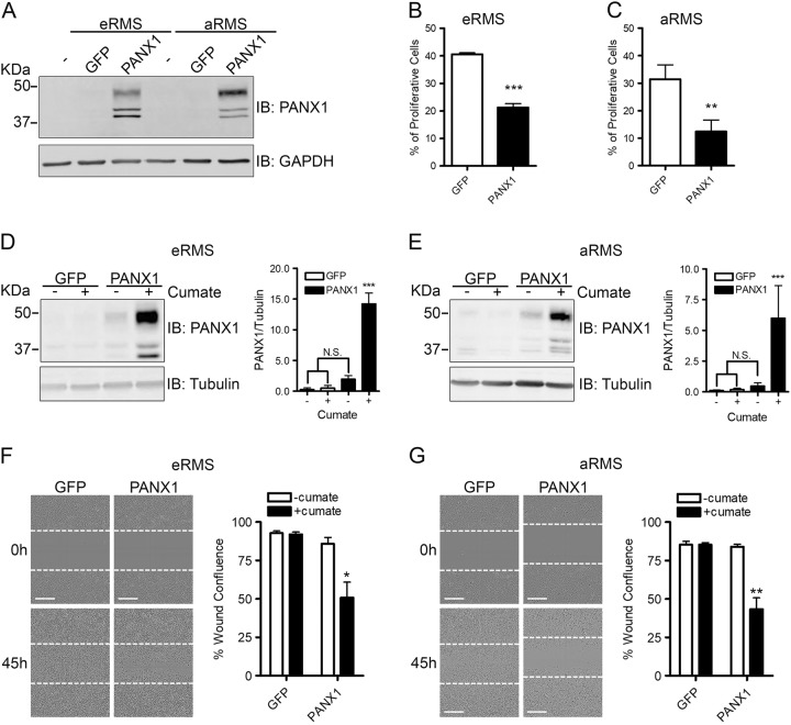
Fig. 2. PANX1 expression inhibits RMS cell proliferation and migration.
eRMS (Rh18) and aRMS (Rh30) cells ectopically expressing PANX1 were analyzed by BrdU incorporation and scratch wound migration assays. a Representative Western blot of eRMS (Rh18) and aRMS (Rh30) cells transiently transfected with PANX1. GAPDH was used as a loading control. BrdU incorporation assay showed a significant reduction of proliferation in eRMS (Rh18) (b) and aRMS (Rh30) (c) cells over-expressing PANX1. ***P < 0.001 and **P < 0.01 compared to GFP. Western blot analysis and quantification of eRMS (Rh18) (d) and aRMS (Rh30) (e) inducible stable cells over-expressing PANX1 after treatment with 30 µg/mL cumate for 24 h. Tubulin was used as loading control. ***P < 0.001 compared to GFP without cumate, GFP with cumate, and PANX1 without cumate. N.S.: not significant. Representative pictures and quantification of stable eRMS (Rh18) (f) and aRMS (Rh30) (g) cells, treated with or without cumate to induce PANX1 over-expression, subjected to scratch wound assay for 45 h. The dotted lines show cell boundaries after initial scratch. The confluence of the wound areas was quantified 45 h post wounding, which showed a significant reduction in cumate-treated PANX1 over-expressing stable eRMS (Rh18) (f) and aRMS (Rh30) (g) compared to their respective controls. **P < 0.01, *P < 0.05 compared to GFP without cumate, GFP with cumate, and PANX1 without cumate. Results are expressed as mean ± s.d. Bars = 300 µm

