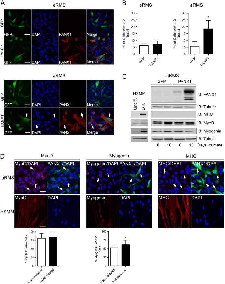
Fig. 3. PANX1 expression does not trigger RMS cell terminal differentiation.
a PANX1 (red) immunofluorescence labeling of eRMS (Rh18) and aRMS (Rh30) transfected with PANX1 or the control vector GFP. While no morphological changes were observed in eRMS cells, a population of PANX1 over-expressing aRMS cells was multinucleated (arrows). Blue = nuclei, bars = 20 µm. b Quantification of multinucleation (% of cells with ≥2 nuclei) of aRMS (Rh30) cells showed a significant increase with PANX1 over-expression. *P < 0.05 compared to GFP. c Cumate-inducible stable aRMS (Rh30) cells were cultured in growth media with 30 µg/mL cumate for 10 days, and analyzed for MHC, MyoD, and myogenin levels. Cells were collected for analysis immediately prior to cumate treatment on Day 0. Undifferentiated and differentiated HSMM were used as myogenic marker controls. Tubulin was used as a loading control. d Representative immunofluorescence images of aRMS (Rh30) cells transfected with PANX1 and labeled with MyoD, myogenin, or MHC (red). Cells transfected with PANX1 (GFP-positive) are shown in green. Differentiated HSMM were used as positive controls. The percentage of mononucleated and multinucleated (arrows) PANX1-expressing cells positive for MyoD, myogenin, and MHC were quantified in a double-blinded manner. *P < 0.05 compared to mononucleated cells. Bars = 20 µm. Results are expressed as mean ± s.d

