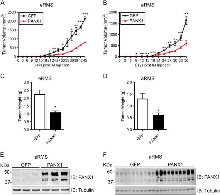
Fig. 5. Expression of PANX1 decreases RMS tumor growth in vivo.
Stable GFP control and PANX1 over-expressing eRMS (Rh18) and aRMS (Rh30) cells were injected orthotopically into the left and right gastrocnemius of the mice (mice randomly assigned, not a blinded method). PANX1 expression was maintained by intraperitoneal injection of cumate every 3 days. PANX1 over-expression in eRMS (Rh18) (a) and aRMS (Rh30) (b) cells led to a significantly reduced growth rate compared to their respective GFP controls. At endpoint, PANX1-expressing eRMS (Rh18) (c) and aRMS (Rh30) (d) xenografts weighted significantly less than the control tumors. Day 0 denotes the day of cell intramuscular (IM) injection. *P < 0.05, **P < 0.01, ***P < 0.001 compared to GFP. Results are expressed as mean ± s.d. Western blots of eRMS (Rh18) (e) and aRMS (Rh30) (f) xenografts demonstrate successful induction of PANX1 by cumate in vivo. Tubulin was used as loading control

