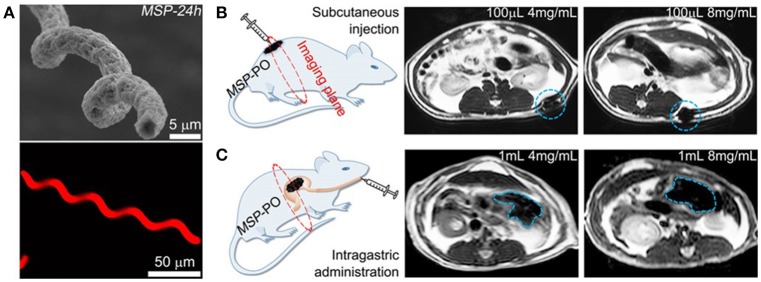
Figure 5.
In vivo imaging of magnetically propelled microrobot. (A) Scanning electron microscopy (SEM) (top) and fluorescence images (bottom) of the helical structured microrobot. Schematic of the target in vivo area and magnetic resonance imaging of microrobots inside rats. Illustrating different microrobot concentrations at the (B) subcutaneous tissues and (C) inside the mouse stomach. Reprinted with permission from Yan et al. (2017). Copyright 2017 The American Association for the Advancement of Science.

