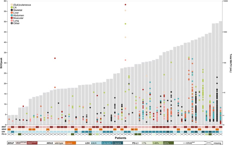
Fig. 1.
Individual tumour lesions (≥ 1 ml) and their SUVpeak displayed per patient. For each patient (x-axis; n = 64), individual tumour lesions are plotted against their SUVpeak (left y-axis). Grey shaded bars represent the patient’s total MATV (right y-axis). The heatmap displays respectively the patient’s LDH level and tumour BRAF and NRAS status and PD-L1 expression. Three patients are not displayed since they only had lesions < 1 ml, which resulted in SUVs, a MATV and TLG of 0. LDH lactate dehydrogenase, LN lymph node, PD-L1 programmed death-ligand 1, ULN upper limit of normal

