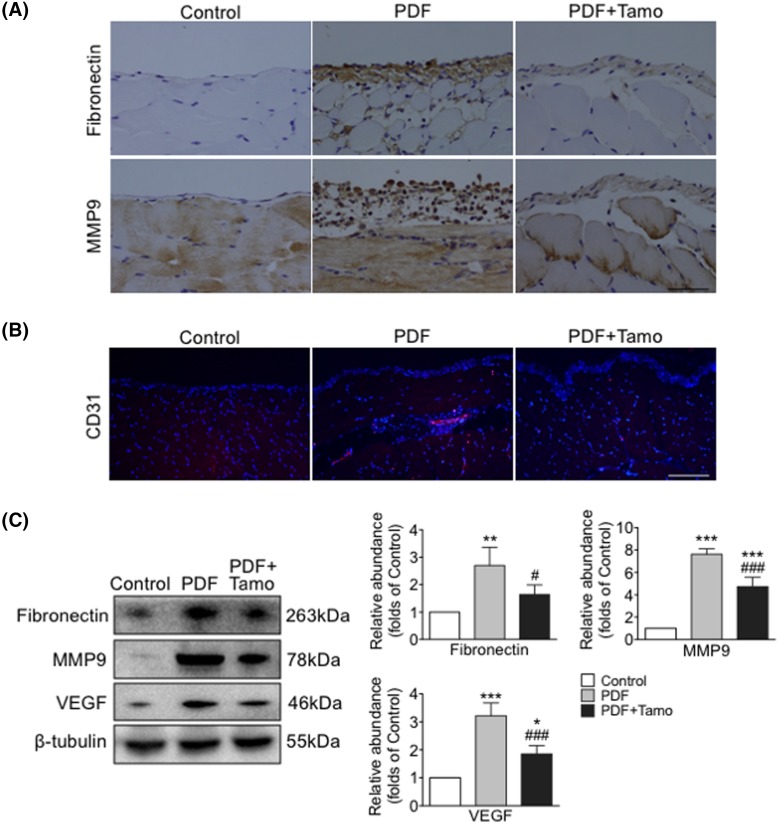
Figure 2. Tamoxifen suppressed up-regulation of fibronectin, MMP9, and angiogenesis following PDF exposure.
Peritoneal tissues collected after 30 days of exposure to 4.25% HG PDF with or without Tamoxifen (Tamo). (A) Paraffin sections incubated with anti-fibronectin or anti-MMP9 antibody (brown). Original magnification: 400×. Bar = 100 μm. (B) Frozen sections were incubated with antibodies against CD31 (red) and nuclei were stained with Hoechst (blue). Original magnification: 400×. Bar = 100 μm. (C) Expression levels of fibronectin, MMP9, and VEGF examined by Western blotting (left panel) and quantitated by densitometry normalized to β-tubulin (right panel) (mean ± S.D., n=5, ***P<0.001 and **P<0.01 compared with control group, ###P<0.001 compared with PDF group; *P<0.05 compared with control group and #P<0.05 compared with PDF group.).

