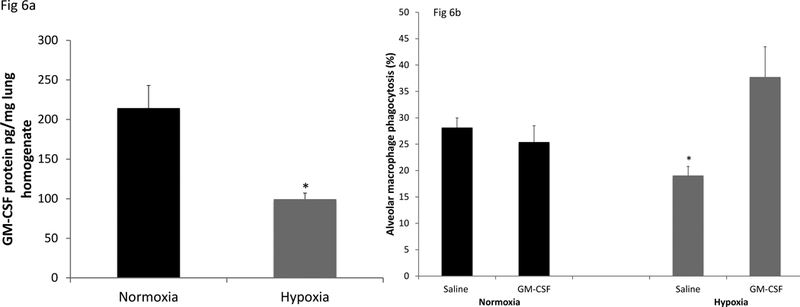
Figure 6. Effect of hypoxia exposure in vivo on lung GM-CSF expression and AM phagocytic activity.

C57BL/6 mice were exposed to room air (21% oxygen) or hypoxia (12% oxygen) for 48h. GM-CSF protein levels in whole lung homogenates were determined (ELISA), normalized to total protein content (Figure 6a). The data are the mean ± SEM; n=5 *p<0.01.Alveolar macrophages were obtained by lung lavage after 48h exposure in 21% or 12% oxygen from mice given saline or GM-CSF (100ng in 40μl saline, IN) at 0 and 24h time of exposure (Figure 6b). Two hours prior to collection of the alveolar macrophages, fluorescent beads (2 ×10⁷ in 40μl saline) were given intra-nasally. The results are expressed as the % macrophages that had taken up beads. 500 macrophages from each mouse were scored and the data are the mean ± SEM; n=5 *p<0.01.
