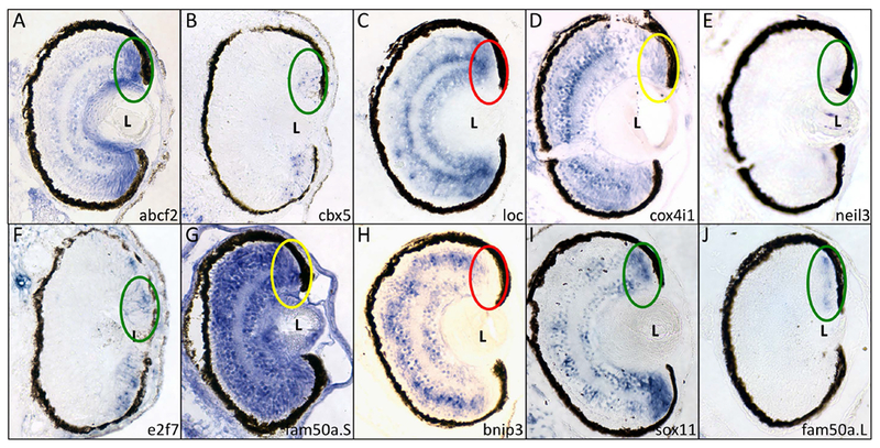
Figure 2 – Six of the genes identified in the microarray screen are expressed in the ciliary marginal zone.

Fixed X. laevis tadpoles (st 41) were paraffinized, sectioned and subjected to in situ hybridization using antisense riboprobes for the 10 genes expressed in the eye (see Figure 1). Genes/riboprobes are identified in the lower left of each panel. Sections are presented with dorsal side towards the top of the figure. The dorsal CMZ is indicated with an oval. Key: green – positive expression in CMZ; red – negative expression in CMZ; yellow – positive expression in CMZ and ubiquitous expression in retina. L – lens.
