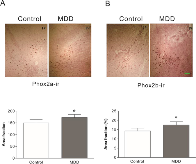
Figure 2:
Phox2a and Phox2b immunoreactivity (ir) in the LC of MDD donors and age-matched psychiatrically normal control donors (N=12 for both). Upper panel: Representative images of Phox2a- and Phox2b-ir in the LC of brains detected by immunocytochemical staining. Lower panel: semi-quantitative analyses of Phox2a-ir and Phox2b-ir in tissue sections. * p<0.05, compared to the control. IV: Fourth ventricle. Scale bar: 250 μm for all images.

