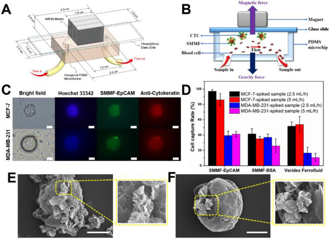
Figure 4.
Microfluidic screening of tumor cells with hierarchical microflower material. (A) Schematic image of our developed microchip for CTCs screening. (B) The cross-section of microchip for showing the working principle in this study. (C) Representative images of captured MCF-7 cell and MDA-MB-231 cell using SMMF-EpCAM. Scale bar = 20 μm. (D) Screening efficiency comparison of SMMF-EpCAM, SMMF-BSA, and Veridex Ferrofluid (from CellSearch™, see details in SI) at different screening flow rates for the tumor cells spiked in whole blood samples. (E) and (F) are the representative SEM images of SMMF-EpCAM-captured MCF-7 cell and MDA-MB-231 cell, respectively. Scale bar = 5 μm.

