
Fig. 10. Representative electron micrographs of mu opioid receptor (MOR) silver intensified gold (SIG) particles in parvalbumin (PARV)-containing interneurons in the dentate gyrus.
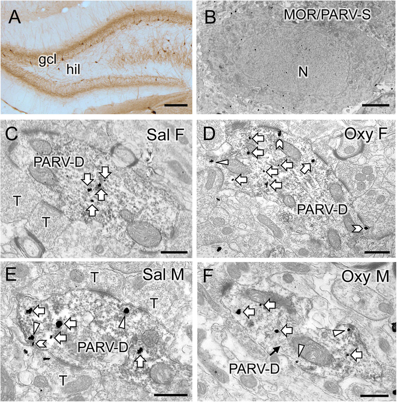
A. Low magnification photomicrograph shows PARV-labeled cells are primarily located in the subgranular region of the hilus (hil) in the dentate gyrus (gcl = granule cell layer). B. Example of somata in the subgranular zone of the hilus dually labeled for MOR-SIG particles and PARV (immunoperoxidase). N, nucleus. C-F. Representative micrographs show the distribution of MOR-SIG particles within peroxidase-labeled PARV dendrites in the dentate gyrus for a Sal-female (C), an Oxy-female (D), a Sal-male (E), and an Oxy-male (D). Examples of on the plasmalemma (chevron), near the plasmalemma (triangle) and cytoplasmic (white arrow) MOR-SIG particles in dendrites are shown. Saline-injected females showed a lower overall density of MOR-SIG particles in PARV-labeled dendrites relative to saline-injected males, but after oxycodone CPP the density of plasmalemmal and total of MOR-SIG particles increased in PARV-labeled dendrites in females only. Scale bars: A = 500 µm; B, C-F = 500 nm.