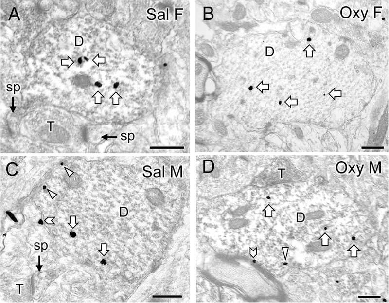
Fig. 4. Representative electron micrographs of delta opioid receptor (DOR) silver-intensified gold (SIG) particles in CA3 pyramidal cell dendrites from saline-injected and oxycodone conditioned place preference rats.

A-D. Electron micrographs show the distribution of DOR-SIG particles within CA3 pyramidal cell dendrites from a Sal-female (A), an Oxy-female (B), a Sal-male (C), and an Oxy-male (D). Examples of on the plasmalemma (chevron), near the plasmalemma (triangle) and cytoplasmic (white arrow) DOR-SIG particles in dendrites are shown. CA3 dendrites often have spines (sp) that are contacted by terminals (T). The Oxy-female has a higher density of DOR-SIG particles in total in CA3 pyramidal dendrites compared to the Sal-female. The Oxy-male has fewer near plasmalemmal DOR-SIG particles in CA3 pyramidal dendrites compared to the Sal-male. Scale bar: 500 nm.
