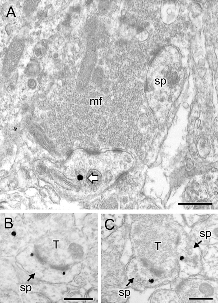
Fig 6. Representative micrographs of delta opioid receptor (DOR)-labeled dendritic spines in CA3.

A. Representative electron micrograph from stratum lucidum of CA3 of an Oxy-male rat shows a DOR silver-intensified gold (SIG) particle in the cytoplasm of a dendritic spine (sp) in contact with a mossy fiber (mf) in the CA3. B, C. Representative electron micrographs from stratum radiatum of CA3 of an Oxy-female rat show DOR-SIG particles near the perforated synapse (B) or in the cytoplasm (C) of two dendritic spines (sp) which are contacted by a terminal (T). Scale bar: 500 nm.
