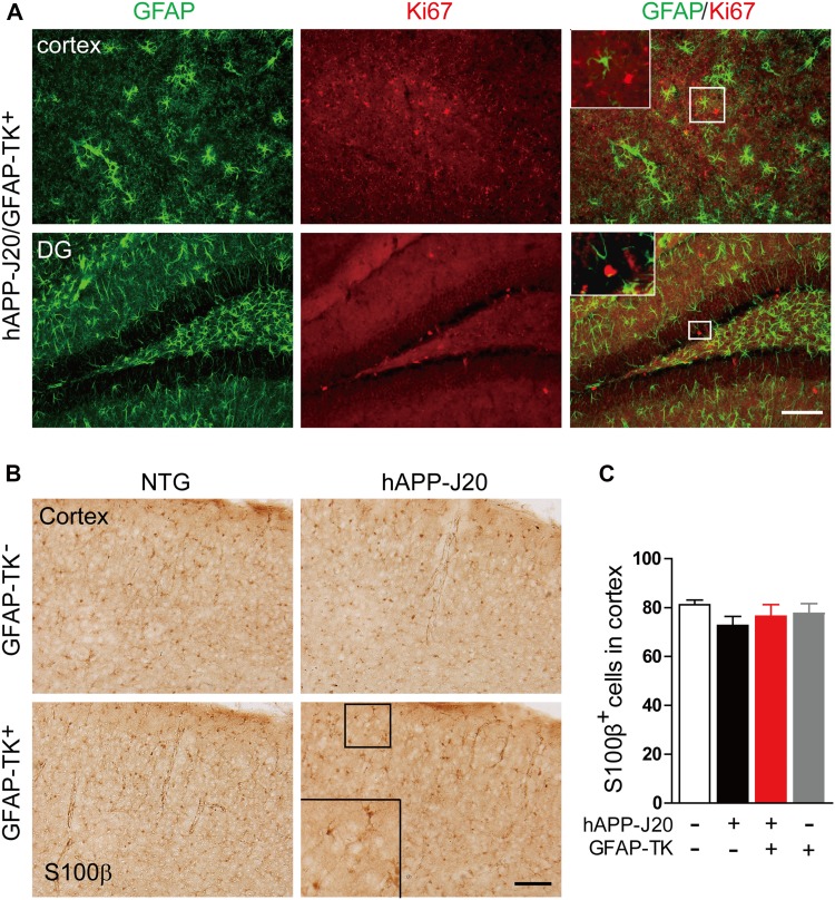
Fig. 2.
Increased expression of GFAP and vimentin in the cortex and hippocampus of hAPP-J20/GFAP-TK+ mice was not due to proliferation of astrocytes. A Photomicrographs of immunofluorescence staining for GFAP and Ki67 in hAPP-J20/GFAP-TK+ mice (scale bar, 100 μm). B Photomicrographs of immunohistochemical staining for S100β in NTG/GFAP-TK−, hAPP-J20/GFAP-TK−, NTG/GFAP-TK+, and hAPP-J20/GFAP-TK+ mice (scale bar, 100 μm). C Quantification of the number of S100β+ cells in the cortex of NTG/GFAP-TK−, hAPP-J20/GFAP-TK−, NTG/GFAP-TK+, and hAPP-J20/GFAP-TK+ mice (n = 20–28 per genotype; data represent mean ± SEM).

