Fig. 6.
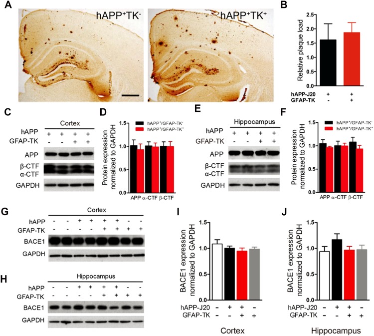
Activation of astrocytes did not affect the amyloid plaque load and the processing of hAPP in hAPP-J20 mice. A Photomicrographs of 3D6 immunostaining of 9-month-old hAPP-J20/GFAP-TK− and hAPP-J20/GFAP-TK+ mice (scale bar, 500 μm). B Percentage of area covered by plaques in the hippocampus (n = 5 mice per genotype). C Protein bands of APP and APP carboxyl-terminal fragments (CTFs) in cortex; GAPDH served as the loading control. D Quantification of APP and APP-CTFs in cortex. E Protein bands of APP and APP-CTFs in hippocampus. F Quantification of the levels of APP and APP-CTFs in the hippocampus of 3-month-old hAPP-J20/GFAP-TK−, and hAPP-J20/GFAP-TK+ mice (n = 8–10 per group) assessed by western blot. G, H Protein bands of BACE1 in cortex and hippocampus. I, J Quantification of the levels of BACE1 in cortex and hippocampus of 3-month-old NTG/GFAP-TK−, hAPP-J20/GFAP-TK−, NTG/GFAP-TK+ and hAPP-J20/GFAP-TK+ mice (n = 4–9 per genotype; data represent mean ± SEM).

