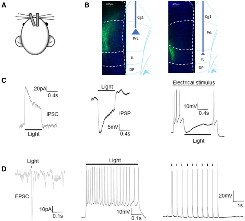
Fig. 4.
Confirmation of optogenetic inhibition or inhibition of neuronal firing in pyramidal neurons. A Schematic of the implanted optic fibers: in the left hemisphere tilted 20°, and vertical on the right side. B EYFP expression in excitatory PL/IL neurons after viral injection. C Examples of yellow light-induced outward current and membrane hyperpolarization in a neuron expressing ArchT. An IPSC (left), IPSP (middle), and inhibition of APs were induced by the yellow light stimulation. D Example of a blue light-evoked EPSC recorded in an EYFP-tagged ChR2-expressing neuron (left). Current clamp recordings under either continuous blue-light stimulation or in response to blue light delivered at interpulse intervals of 0.5 s. The pulse-locked neuronal firing was induced by the blue light, confirming the expression and function of ChR2 in the pyramidal neuron (middle and left).

