Fig. 1.
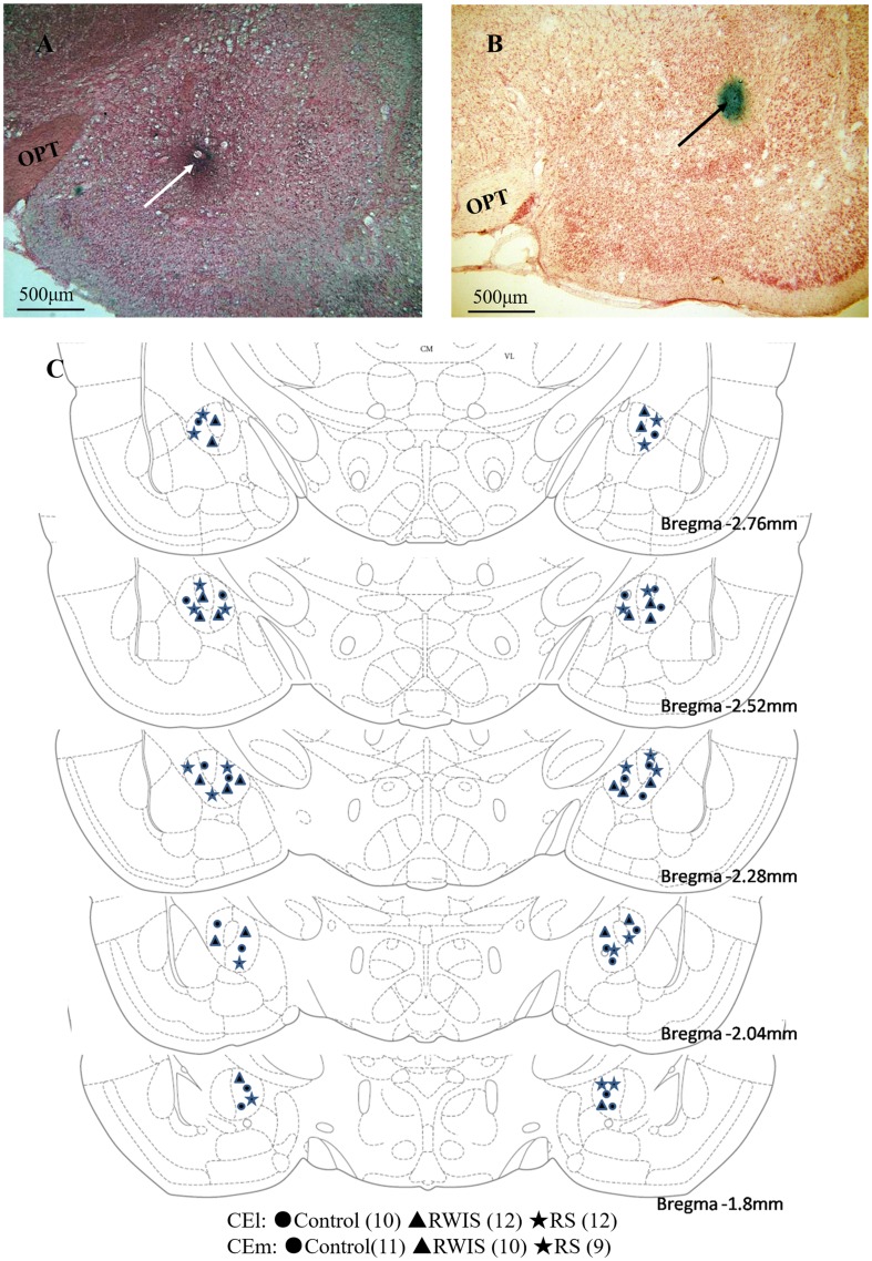
Histological verification of recording sites. A–B Two representative examples of coronal sections stained with neutral red, photographed at low magnification, demonstrating the positions of microelectrode tips in the medial part of the central nucleus of the amygdala (white arrow) and the lateral part of the central nucleus of the amygdala (black arrow). C Schematic reconstructions of recording locations in the control (circles), RWIS (triangle), and RS groups (stars). Distances of coronal sections posterior to bregma are according to the rat brain atlas of Paxinos and Watson [23]. OPT, optic tract.

