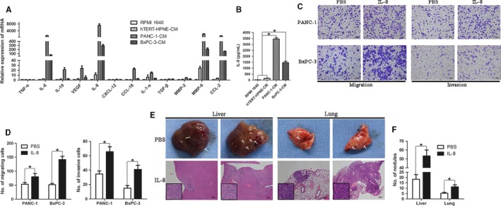
Figure 3.

Tumor‐driven like macrophages secreted IL‐8 to enhance pancreatic ductal adenocarcinoma cells motility. A, THP‐1 monocytes were treated with different conditioned media (CM; RPMI 1640, hTERT‐HPNE‐CM, PANC‐1‐CM, or BxPC‐3‐CM) for 48 h. The RNAs was extracted from different cells by Trizol reagent, and then, cDNA was synthesized.The mRNA expression of cytokines in different CM‐treated THP‐1 cells was assayed by qRT‐PCR. B, IL‐8 protein expression level in supernatant of different CM‐treated THP‐1 cells was determined by ELISA. C, PANC‐1 and BxPC‐3 cells (2 × 10⁴/well) treated with 100 ng/mL IL‐8 or PBS were evaluated their migratory and invasive abilities by Transwell assay. Magnification: 100×; Scale bars: 100 μm. D, Histogram of average numbers of penetrated cells per microscopic field. E, A single‐cell suspension of PANC‐1 cells (2 × 10⁶ cells) in 0.2 mL of PBS was injected into tail vein of nude mice, and exogenous recombinant human IL‐8 (1 μg per mouse and PBS as control) was injected via peritoneal cavity. On day 21, the mice were sacrificed and dissected. Gross metastatic lesion in liver and lung and their H&E staining were showed in the images. Magnification: 40×; Scale bars: 200 μm. F, Graphic representation of liver and lung metastatic lesions per slide under microscopy. *P < 0.05
