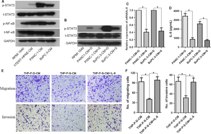
Figure 4.

Tumor‐driven like macrophages secreted IL‐8 through activating STAT3 pathway. A, THP‐1 monocytes were cultured with corresponding conditioned media (CM) for 48 h and analyzed for levels of phosphorylated STAT3 (p‐STAT3) and NF‐κB (p‐NF‐κB). B, The effective inhibition of p‐STAT3 by STATTIC was confirmed by Western blotting. C, The mRNA expression level of IL‐8 in tumor‐driven like macrophages at the presence of DMSO or STATTIC (15 µmol/L, dissolved in DMSO) was evaluated by qRT‐PCR. D, IL‐8 protein expression level in tumor‐driven like macrophages at the presence of DMSO or STATTIC (15 µmol/L) was evaluated by ELISA. E, THP‐1 monocytes were treated with 15 µmol/L STATTIC for 1 h and corresponding 50% v/v CM of PANC‐1 cells for 48 h, and then, the CM was collected. Migratory and invasive abilities of PANC‐1 under different conditions were determined by Transwell assay. F, Histogram of average numbers of penetrated cells per microscopic field. Magnification: 100×; Scale bars: 100 μm. *P < 0.05. THP‐P‐D‐CM: CM produced by THP‐1 cells cultured in the presence of 50% v/v PANC‐1‐CM and DMSO; THP‐P‐S‐CM: CM produced by THP‐1 cells cultured in the presence of 50% v/v PANC‐1‐CM and STATTIC
