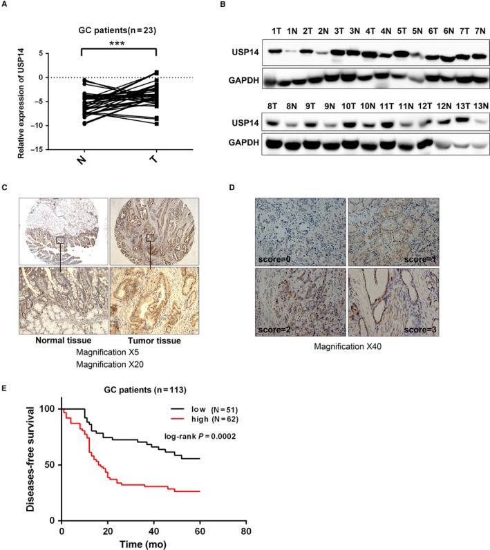
Figure 2.

USP14 expression level was increased in GC samples and an independent prognostic marker for DFS in GC. A, Elevated USP14 expression was observed in our own GC samples (N = 23). B, Similarly, USP14 protein level was measured via Western blotting in 13 paired GC and adjacent normal specimens, showing that USP14 protein level were increased in most of the detected cancer tissues (9/13). C, Immunohistochemistry assay showed stronger cytosolic staining intensities of USP14 in GC samples compared with matched normal counterparts (10/15). D, The staining intensities of USP14 in GC samples were scored as 0, 1, 2, and 3, among which 3 indicated the strongest intensity. The representative four staining intensities are shown here. The expression levels of USP14 were determined via multiplying intensities by positive areas. The low expressions of USP14 were 0‐3 points, while the high ones were 4‐12 points. E, GC patients (N = 113) with high USP14 expression levels have an unfavorable DFS (P = 0.0002)
