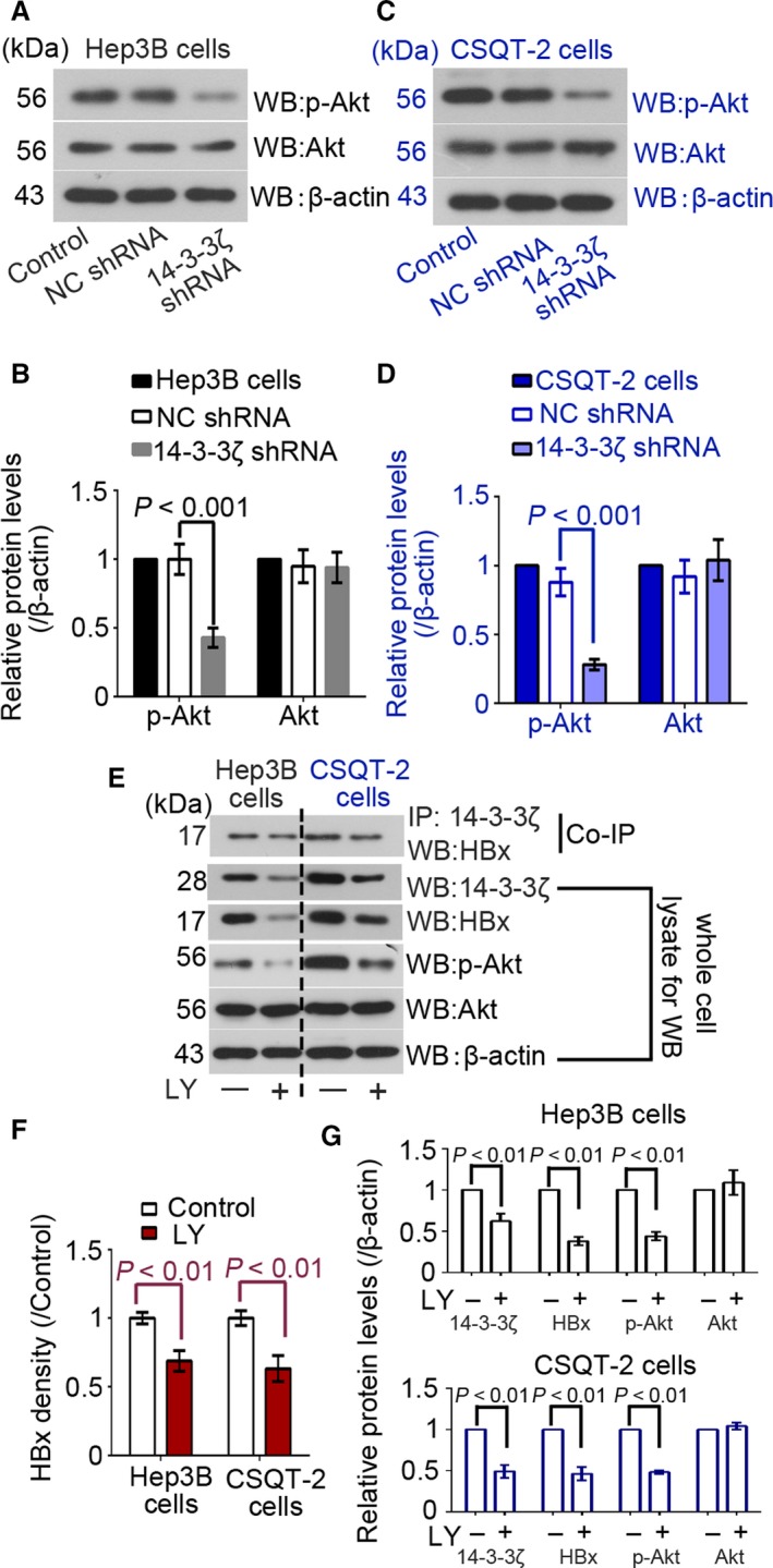
Figure 2.

Inhibition of Akt pathway suppresses the interaction between 14‐3‐3ζ and pSer31‐HBx. Hep 3B and CSQT‐2 cells were infected with lentiviral particles containing 14‐3‐3ζ shRNA or NC shRNA. The levels of p‐Akt and total Akt in Hep 3B (A and B) and CSQT‐2 cells (C and D) were determined with Western blotting analysis with β‐actin as an endogenous reference (n = 3). Hep 3B and CSQT‐2 cells were treated with or without LY294002 (20 μmol/L) for 24 h. (E and F) IP with anti‐14‐3‐3ζ antibody and Western blotting assay with anti‐HBx antibody were performed in Hep 3B and CSQT‐2 cells. (E and G) The levels of 14‐3‐3ζ, HBx and phosphorylated‐Akt (p‐Akt) and total Akt were determined with Western blotting analysis with β‐actin as an endogenous reference (n = 3).
