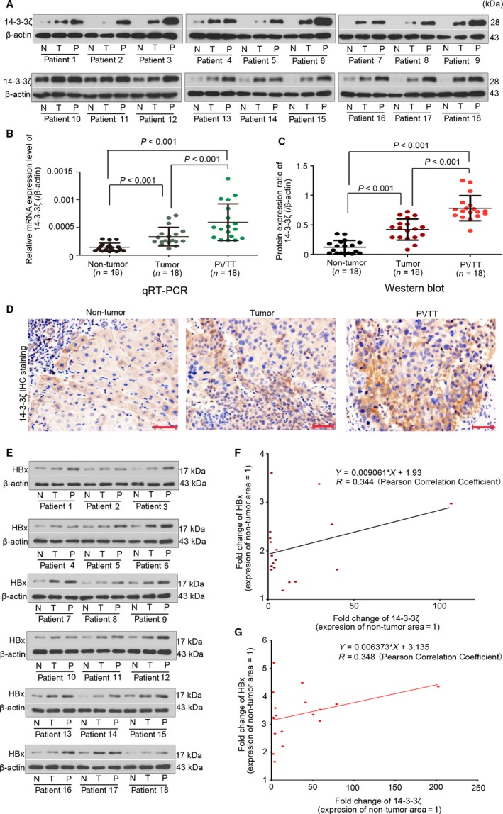
Figure 5.

14‐3‐3ζ correlates with HBx to be elevated in primary and metastatic tumor tissue of HCC patients. The 14‐3‐3ζ protein expression in nontumor, primary tumor, and PVTT tissue (n = 18 pairs) was analyzed with (A,C) Western blotting analysis (with β‐actin as an endogenous reference) and (D) IHC staining (Bars = 50 μm). (B) Its mRNA expression was determined with qRT‐PCR analysis. The protein levels of HBx were also determined via Western blotting analysis with β‐actin as an endogenous reference (E). Fold changes of 14‐3‐3ζ and HBx were calculated by comparing their protein expression levels of the primary tumor (F) and PVTT tissues (G) to that of the nontumor tissue and then analyzed with Pearson correlation.
