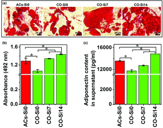
Figure 6.

Oil Red O staining and adiponectin secretion of monocultured cells and cocultured cells treated with or without Si ions. a) Oil Red O staining of cells showed that Si ions stimulated the increase of lipid accumulation in the coculture system and inhibited the dedifferentiation of cocultured adipocytes. Lipid droplets were stained in red. Scale bar, 20 µm. b) Quantitative analysis of Oil Red O staining (n = 3 for both the groups; *p < 0.05). c) Quantitative ELISA analysis showed that Si ions enhanced the adiponectin secretion of cocultured cells in culture medium (n = 3 for both the groups; *p < 0.05). ACs‐Si0: HBMSCs cultured in adipogenic differentiation medium without Si ions for 12 days for the development of the adipocytes' (ACs) phenotype; CO‐Si0, CO‐Si7, CO‐Si14: HBMSCs first cultured in adipogenic differentiation medium with different concentrations of Si ions (0, 7, and 14 µg mL−1) for 12 days for adipogenic differentiation, and then cocultured with HUVECs containing different concentrations of Si ions (0, 7, and 14 µg mL−1) for 3 days.
