Figure 7.

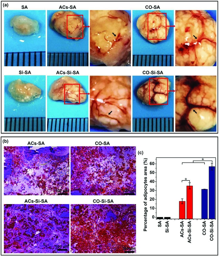
Tissue appearance and Oil Red O staining of engineered adipose tissue in a nude mice subcutaneous implant model by combining the monocultured cells or cocultured cells in calcium silicate/alginate composite hydrogel with the ability to release bioactive Si ions. a) Appearance of engineered adipose tissue showed that Si ions stimulated adipose tissue growth and vascular network (black arrows) formation. High magnification corresponded to boxed area (red) in the low magnification images. b) Oil Red O staining of implants showed that Si ions promoted adipose tissue regeneration in vivo. Lipid droplets were stained in red and nucleus were counterstained in blue with hematoxylin. Scale bar, 100 µm. c) Quantitative analysis of Oil Red O staining of the implants indicated that Si ions stimulated adipose tissue formation (n = 5 for both the groups; *p < 0.05). SA: hydrogels without cells; Si‐SA: Si ion–released hydrogels without cells; ACs‐SA: hydrogels with monocultured adipocytes; ACs‐Si‐SA: Si ion–released hydrogels with monocultured adipocytes; CO‐SA: hydrogels with cocultured adipocytes and HUVECs; CO‐Si‐SA: Si ion–released hydrogels with cocultured adipocytes and HUVECs.
