Figure 6.

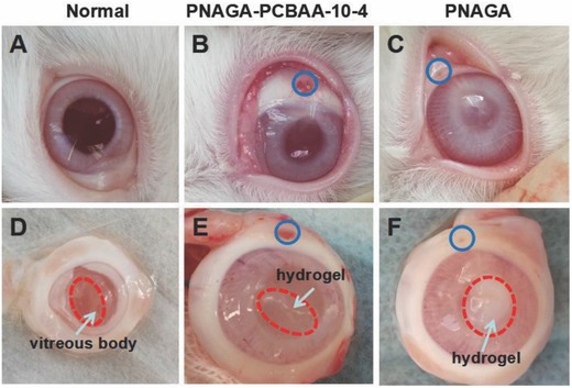
Appearance of A) normal group eye, the eye implanted with B) PNAGA‐10‐4 hydrogel, and C) the eye implanted with PNAGA hydrogel. Incised eyes showing the states of D) normal vitreous body, E) PNAGA‐PCBAA‐10‐4 hydrogel, and F) PNAGA hydrogel implanted in the vitreous cavity for 4 weeks. Blue circle indicated the injection hole.
