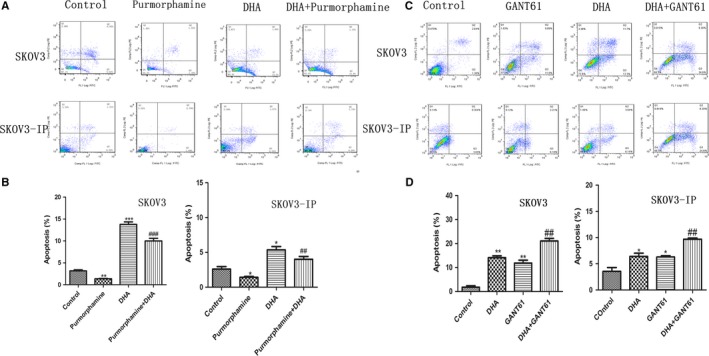
Figure 6.

The Hh signaling pathway is involved in DHA‐induced apoptosis. A and B, SKOV3 and SKOV3‐IP cells were treated with 1.5 µM of purmorphamine, DHA, or combined DHA and purmorphamine for 48 h. C and D, SKOV3 and SKOV3‐IP cells were treated with 20 µM GANT61, DHA, or combined DHA and GANT61 for 48 h. The concentration of DHA in SKOV3 and SKOV3‐IP was 80 and 40 µM, respectively, and the control group was treated with DMSO. Apoptosis was analyzed by flow cytometry. All experiments were repeated three times. *P < 0.05 and **P < 0.01, vs the control group. # P < 0.05, ## P < 0.01 and ### P < 0.001, vs the purmorphamine and GANT61 groups
