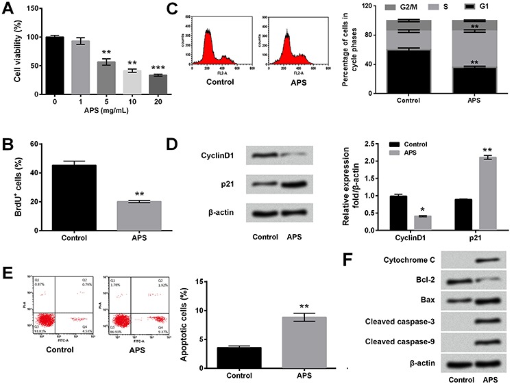
Figure 1. Astragalus polysaccharides (APS) reduced proliferation and induced apoptosis of MG63 cells. MG63 cells were treated by 1-20 mg/mL APS. Non-treated cells acted as control. A, Cell viability was detected by CCK-8 assay. B, Percentage of BrdU positive cells was measured by BrdU incorporation assay. C, Cell cycle distribution was assessed using PI staining and flow cytometry. D, Expression levels of proliferation-associated proteins were evaluated by western blot analysis. E, Percentage of apoptotic cells was determined by FITC Annexin V/Dead Cell Apoptosis kit and flow cytometry. F, Expression levels of apoptosis-associated proteins were evaluated by western blot analysis. Data are reported as means±SE of three independent experiments. *P<0.05; **P<0.01; ***P<0.001 (ANOVA or t-test).

