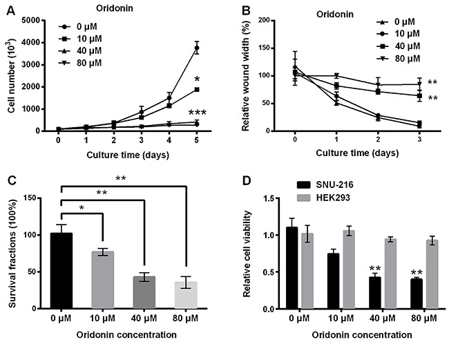
Figure 1. Oridonin inhibited the growth, migration, and survivability of SNU-216 cells. Cells were treated with a series of oridonin (0, 10, 40 and 80 μM) for indicated days. A, Viable cell number was measured by trypan blue dye staining. B, Cell migration was assessed by wound healing assay. After treatment with oridonin for 24 h, the survival fraction of cells was detected by clonogenic assay (C), and cell viability of SNU-216 cells and HEK293 cells was measured by CCK-8 assay (D). Data are reported as means±SD. *P<0.05, **P<0.01, ***P<0.001 compared to 0 μM (ANOVA).

