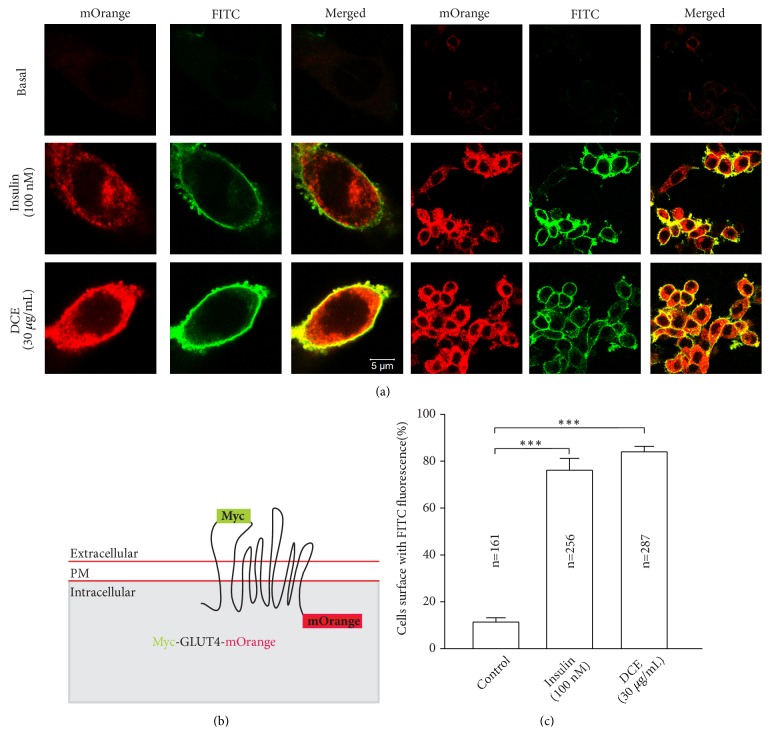
Figure 6.
DCE increased the GLUT4 fusion with PM. (a) L6 cells were transfected with plasmid GV348-myc-GLUT4-mOrange encoding a mOrange fusion protein with myc epitope-tagged GLUT4 (myc-GLUT4-mOrange). Cells were stimulated with 100 nM insulin or 30 μg/mL DCE for 30 minutes, respectively. Then fixed and hybridized with specific immunofluorescent antibodies, FITC fluorescence was measured. Scale: 5 μm. (b) GLUT4 fusion protein for detection of GLUT4 translocation and PM surface exposure. (c) Statistical percentage of FITC positive cells in mOrange positive cells. Summary results are from three independent experiments. ∗∗∗P < 0.001.

