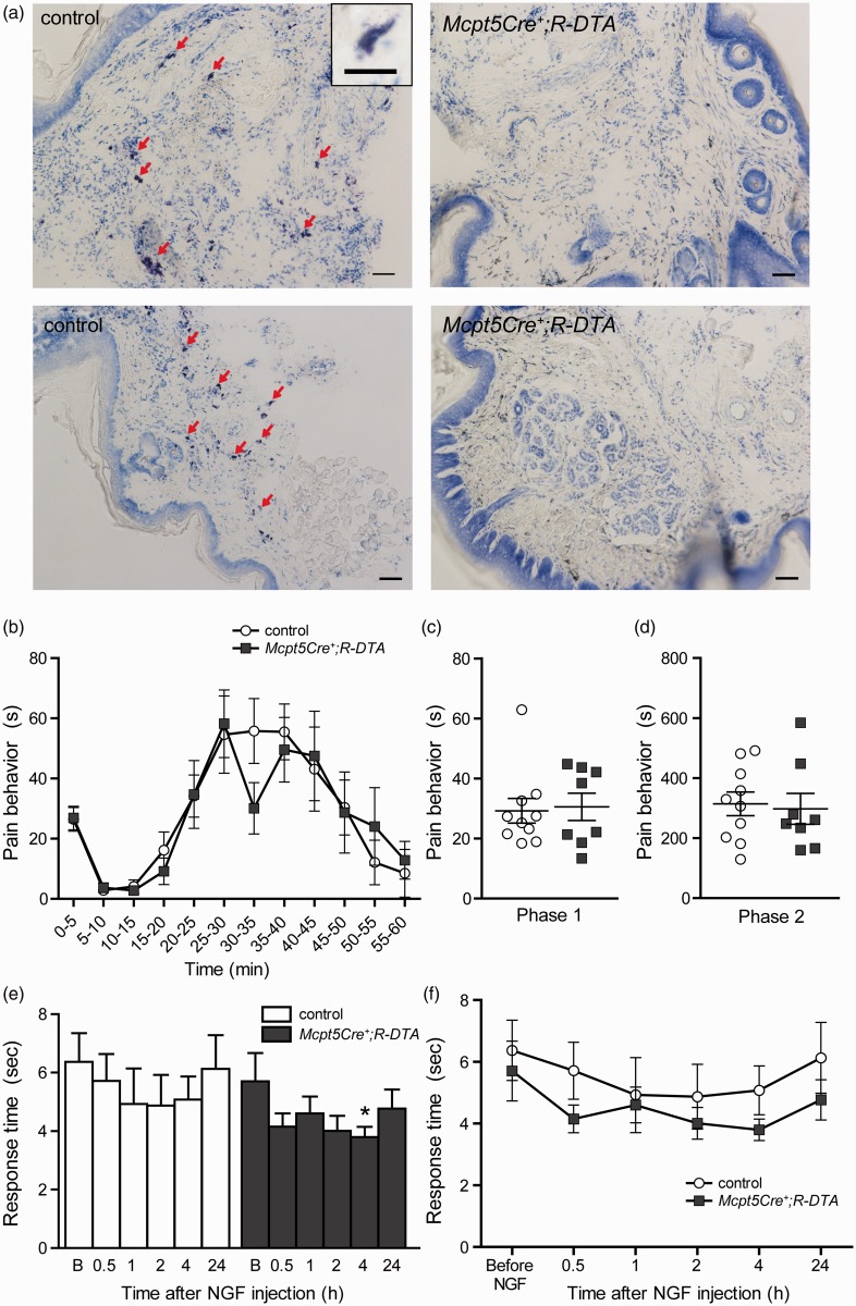
Figure 2.
Mast cell-deficient mice do not differ from controls in the formalin test or in heat hypersensitivity induced by NGF. (a) Toluidine blue-stained cryo sections of hind paws. Plantar surfaces of hind paws of naïve Mcpt5Cre-;R-DTA (controls, n = 2) and Mcpt5Cre+;R-DTA (n = 2) were fixed with PFA, embedded in OCT compound, cut into 12-µm-thick sections, and subsequently stained with toluidine blue. Red arrows point to metachromatically stained mast cells in control tissue, which are absent in tissue of Mcpt5Cre+;R-DTA mice. 10× objective, scale bar = 50 µm. Inset represents metachromatically stained mast cell, 20× objective, scale bar = 20 µm. (b) The pain behavior of mast cell-deficient mice (Mcpt5Cre+;R-DTA, n = 8) does not differ from controls (n = 10) over the time course of the formalin test.(Continued)Slightly lower responses were observed at 30–35 min, but the difference was not significant (Mann–Whitney: p = 0.10). (c) and (d) When total pain behavior in Phase 1 (0–10 min) or in the inflammatory Phase 2 (10–60 min) was analyzed, no differences were observed between genotypes. (e–f) Hargreaves test coupled with NGF injection on Mcpt5Cre+;R-DTA mice (n = 7), and controls (n = 6). (e) The baseline Hargreaves values before NGF injection (“B” on the x-axis) did not differ between the two genotypes (Student’s t test: p = 0.64). After NGF injection, only Mcpt5Cre+;R-DTA mice developed heat hypersensitivity compared with their baseline values, at 4-h post-injection (one-way repeated measurement ANOVA: p < 0.05). (f) Despite the absence of significant hypersensitivity in the controls, there was no difference observed between the two genotypes at any time point before or after the injection (Student’s t test: p > 0.05). All data are presented as mean ± SEM. NGF: nerve growth factor.

